Animal Tissues – Epithelium, Connective Tissues
Animal Tissues – Epithelial Tissue: Simple Epithelium and Compound Epithelium, Connective Tissue, Muscular Tissue and Nervous Tissue.
General Science: NCERT Science Textbooks Class 6-12.
Animal Tissues
- Blood and muscles are both examples of tissues found in our body. On the basis of the functions they perform we can think of different types of animal tissues, such as epithelial tissue, connective tissue, muscular tissue and nervous tissue.
- Blood is a type of connective tissue, and muscle forms muscular tissue.
Epithelial Tissue
- The covering or protective tissues in the animal body are epithelial tissues.
- Epithelium covers most organs and cavities within the body.
- It also forms a barrier to keep different body systems separate.
- The skin, the lining of the mouth, the lining of blood vessels, lung alveoli and kidney tubules are all made of epithelial tissue.
- Epithelial tissue cells are tightly packed and form a continuous sheet.
- They have only a small amount of cementing material between them and almost no intercellular spaces.
- Obviously, anything entering or leaving the body must cross at least one layer of epithelium.
- As a result cells of various epithelia play an important role in regulating the exchange of materials between the body and the external environment and also between different parts of the body.
- Regardless of the type, all epithelium is usually separated from the underlying tissue by an extracellular fibrous basement membrane.
- There are two types of epithelial tissues namely simple epithelium and compound epithelium.
Simple Epithelium
- Simple epithelium is composed of a single layer of cells and functions as a lining for body cavities, ducts, and tubes.
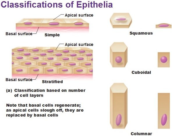
Squamous Epithelium
- The squamous epithelium is made of a single thin layer of flattened cells with irregular boundaries.
- They are found in the walls of blood vessels and air sacs of lungs and are involved in functions like forming a diffusion boundary.
- Different epithelia show differing structures that correlate with their unique functions. For example, in cells lining blood vessels or lung alveoli, where transportation of substances occurs through a selectively Permeable surface, there is a simple flat kind of epithelium. This is called the simple Squamous epithelium.
- Simple squamous epithelial cells are extremely thin and flat and form a delicate lining.
- The oesophagus and the lining of the mouth are also covered with squamous epithelium.
Stratified Squamous Epithelium
- The skin, which protects the body, is made of squamous epithelium.
- Skin epithelial cells are arranged in many layers to prevent wear and tear.
- Since they are arranged in a pattern of layers, the epithelium is called stratified squamous epithelium.
Ciliated Columnar Epithelium
- The columnar epithelium is composed of a single layer of tall and slender cells. Their nuclei are located at the base.
- Where absorption and secretion occur, as in the inner lining of the intestine, tall epithelial cells are present.
- In the respiratory tract, the columnar epithelial tissue also has cilia, which are hair-like projections on the outer surfaces of epithelial cells. These cilia can move, and their movement pushes the mucus forward to clear it. This type of epithelium is thus ciliated columnar epithelium.
- They are mainly present in the inner surface of hollow organs like bronchioles and fallopian tubes.
Cuboidal Epithelium
- The cuboidal epithelium is composed of a single layer of cube-like cells. This is commonly found in ducts of glands and tubular parts of nephrons in kidneys and its main functions are secretion and absorption.
- Cuboidal epithelium (with cube-shaped cells) forms the lining of kidney tubules and ducts of salivary glands, where it provides mechanical support.
Glandular Epithelium
- Epithelial cells often acquire additional specialization as gland cells, which can secrete substances at the epithelial surface.
- Sometimes a portion of the epithelial tissue folds inward, and a multicellular gland is formed. This is glandular epithelium.
- Some of the columnar or cuboidal cells get specialized for secretion and are called glandular epithelium. They are mainly of two types: unicellular, consisting of isolated glandular cells (goblet cells of the alimentary canal), and multicellular, consisting of cluster of cells (salivary gland).
- On the basis of the mode of pouring of their secretions, glands are divided into two categories namely EXOCRINE and ENDOCRINE.
- Exocrine glands secrete mucus, saliva, earwax, oil, milk, digestive enzymes and other cell products. These products are released through ducts or tubes.
- In contrast, endocrine glands do not have ducts. Their products called hormones are secreted directly into the fluid bathing the gland.
Compound Epithelium
- The compound epithelium consists of two or more cell layers and has protective function as it does in our skin.
- Compound epithelium is made of more than one layer (multi-layered) of cells and thus has a limited role in secretion and absorption. Their main function is to provide protection against chemical and mechanical stresses.
- They cover the dry surface of the skin, the moist surface of buccal cavity, pharynx, inner lining of ducts of salivary glands and of pancreatic ducts.
- All cells in epithelium are held together with little intercellular material. In nearly all animal tissues, specialized junctions provide both structural and functional links between its individual cells.
- Three types of cell junctions are found in the epithelium and other tissues. These are called as tight, adhering and gap junctions.
- Tight junctions help to stop substances from leaking across a tissue. Adhering junctions perform cementing to keep neighboring cells together. Gap junctions facilitate the cells to communicate with each other by connecting the cytoplasm of adjoining cells, for rapid transfer of ions, small molecules and sometimes big molecules.
Connective Tissue
- Connective tissues are most abundant and widely distributed in the body of complex animals. They are named connective tissues because of their special function of linking and supporting other tissues/organs of the body.
- They range from soft connective tissues to specialized types, which include cartilage, bone, adipose, and blood.
- In all connective tissues except blood, the cells secrete fibres of structural proteins called collagen or elastin.
- The fibres provide strength, elasticity and flexibility to the tissue. These cells also secrete modified polysaccharides, which accumulate between cells and fibres and act as matrix (ground substance).
- Connective tissues are classified into three types: (i) Loose connective tissue, (ii) Dense connective tissue and (iii) Specialized connective tissue.
Loose Connective Tissue
- Loose connective tissue has cells and fibres loosely arranged in a semi-fluid ground substance, for example, areolar tissue present beneath the skin.
- Often it serves as a support framework for epithelium. It contains fibroblasts (cells that produce and secrete fibres), macrophages [a large phagocytic cell found in stationary form in the tissues or as a mobile white blood cell, especially at sites of infection] and mast cells [a cell found in connective tissue and releasing histamine and other substances during inflammatory and allergic reactions].
- Adipose tissue is a type of loose connective tissue located mainly beneath the skin. The cells of this tissue are specialized to store fats. The excess of nutrients which are not used immediately are converted into fats and are stored in this tissue.
Dense Connective Tissue
- Fibres and fibroblasts are compactly packed in the dense connective tissues. Orientation of fibres show a regular or irregular pattern and are called dense regular and dense irregular tissues.
- In the dense regular connective tissues, the collagen fibres are present in rows between many parallel bundles of fibres. Tendons, which attach skeletal muscles to bones and ligaments which attach one bone to another are examples of this tissue.
- Dense irregular connective tissue has fibroblasts and many fibres (mostly collagen) that are oriented differently. This tissue is present in the skin.
Specialized Connective Tissue – Cartilage, Bones, Blood, Areolar
- Cartilage, bones and blood are various types of specialized connective tissues.
- The intercellular material of cartilage is solid and pliable and resists compression. Cells of this tissue (chondrocytes) are enclosed in small cavities within the matrix secreted by them.
- Most of the cartilages in vertebrate embryos are replaced by bones in adults. Cartilage is present in the tip of nose, outer ear joints, trachea, larynx, between adjacent bones of the vertebral column, limbs and hands in adults.
- Bone cells are embedded in a hard matrix that is composed of calcium and phosphorus compounds.
- Bones have a hard and non-pliable ground substance rich in calcium salts and collagen fibres which give bone its strength. It is the main tissue that provides structural frame to the body. Bones support and protect softer tissues and organs.
- The bone cells (osteocytes) are present in the spaces called lacunae. The bone marrow in some bones is the site of production of blood cells.
- Two bones can be connected to each other by another type of connective tissue called the ligament. This tissue is very elastic. It has considerable strength. Ligaments contain very little matrix. Tendons connect bones to muscles and are another type of connective tissue. Tendons are fibrous tissue with great strength but limited flexibility.
- Blood is a fluid connective tissue containing plasma, red blood cells (RBC), white blood cells (WBC) and platelets. It is the main circulating fluid that helps in the transport of various substances.
- Areolar connective tissue is found between the skin and muscles, around blood vessels and nerves and in the bone marrow.
- It fills the space inside the organs, supports internal organs and helps in repair of tissues.
Muscular Tissue
- Each muscle is made of many long, cylindrical fibres arranged in parallel arrays. These fibres are composed of numerous fine fibrils, called myofibrils.
- Muscle fibres contract (shorten) in response to stimulation, then relax (lengthen) and return to their uncontracted state in a coordinated fashion. Muscles contain special proteins called contractile proteins, which contract and relax to cause movement.
- Muscles are of three types, skeletal, smooth, and cardiac.
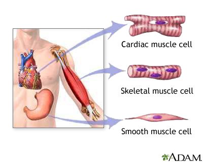
Skeletal Muscle Tissue – Voluntary Muscles
- We can move some muscles by conscious will. Such muscles are called voluntary muscles.
- These muscles are also called skeletal muscles as they are mostly attached to bones and help in body movement.
- Under the microscope, these muscles show alternate light and dark bands or striations. As a result, they are also called striated muscles. The cells of this tissue are long, cylindrical, unbranched and multinucleate (having many nuclei).
- Skeletal muscle tissue is closely attached to skeletal bones. In a typical muscle such as the biceps, striated (striped) skeletal muscle fibres are bundled together in a parallel fashion. A sheath of tough connective tissue encloses several bundles of such muscle fibres.
Smooth Muscle Tissue – Involuntary Muscles
- The movement of food in the alimentary canal or the contraction and relaxation of blood vessels are involuntary movements. We cannot really start them or stop them simply by wanting to do so! Smooth muscles or involuntary muscles control such movements.
- They are also found in the iris of the eye, in ureters and in the bronchi of the lungs.
- The cells are long with pointed ends (spindle-shaped) and uninucleate (having a single nucleus). They are also called unstriated muscles.
- The smooth muscle fibres taper at both ends (fusiform, spindle-shaped) and do not show striations. Cell junctions hold them together and they are bundled together in a connective tissue sheath. The wall of internal organs such as the blood vessels, stomach and intestine contains this type of muscle tissue.
Cardiac Muscle Tissue – Involuntary Muscles
- The muscles of the heart show rhythmic contraction and relaxation throughout life. These involuntary muscles are called cardiac muscles. Heart muscle cells are cylindrical, branched and uninucleate.
- Cardiac muscle tissue is a contractile tissue present only in the heart. Cell junctions fuse the plasma membranes of cardiac muscle cells and make them stick together. Communication junctions (intercalated discs) at some fusion points allow the cells to contract as a unit, i.e., when one cell receives a signal to contract, its neighbors are also stimulated to contract.
Nervous Tissue
- Neural tissue exerts the greatest control over the body’s responsiveness to changing conditions.
- Neurons, the unit of neural system are excitable cells. The neuroglial cell which constitute the rest of the neural system protect and support neurons.
- Neuroglia make up more than one-half the volume of neural tissue in our body.
- When a neuron is suitably stimulated, an electrical disturbance is generated which swiftly travels along its plasma membrane.
- Arrival of the disturbance at the neuron’s endings, or output zone, triggers events that may cause stimulation or inhibition of adjacent neurons and other cells.
- All cells possess the ability to respond to stimuli. However, cells of the nervous tissue are highly specialized for being stimulated and then transmitting the stimulus very rapidly from one place to another within the body.
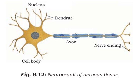
- The brain, spinal cord and nerves are all composed of the nervous tissue. The cells of this tissue are called nerve cells or neurons.
- A neuron consists of a cell body with a nucleus and cytoplasm, from which long thin hair-like parts arise. Usually each neuron has a single long part, called the axon, and many short, branched parts called dendrites.
- An individual nerve cell may be up to a metre long. Many nerve fibres bound together by connective tissue make up a nerve.
- Nerve impulses allow us to move our muscles when we want to. The functional Combination of nerve and muscle tissue is fundamental to most animals. This combination enables animals to move rapidly in response to stimuli.