
Human Digestive System | Digestive Glands
Human Digestive System – Digestive Glands, Enzyme Action in Stomach, Enzyme Action in Small Intestine, Absorption of Digested Products, Disorders of Digestive System.
Human Digestive System
- Biomacromolecules (carbohydrates, proteins etc.) in food cannot be utilized by our body in their original form.
- They have to be broken down and converted into simple substances (glucose, amino acids etc.) in the digestive system.
- During the digestion process, Biomacromolecules like
- carbohydrates get broken into simple sugars such as glucose,
- fats into fatty acids and glycerol,
- proteins into amino acids.
- This process of conversion of complex food substances to simple absorbable forms is called digestion.
Basics:
- Cell Organelles | Plant Cell vs. Animal Cell
- Carbohydrates | Monosaccharides | Polysaccharides
- Proteins | Amino Acids | Enzymes
- Vitamins and Minerals – Deficiency Diseases
- Fats | Healthy Fats and Unhealthy Fats
- Animal Tissues – Epithelium, Connective Tissues
Alimentary Canal
- The food passes through a continuous canal called alimentary canal. The canal can be divided into various compartments: (1) the buccal cavity, (2) foodpipe or oesophagus, (3) stomach, (4) small intestine, (5) large intestine ending in the rectum and (6) the anus.
- The activities of the gastro-intestinal tract [alimentary canal] are under neural and hormonal control for proper coordination of different parts.
- The sight, smell and/or the presence of food in the oral cavity can stimulate the secretion of saliva.
- Gastric and intestinal secretions are also, similarly, stimulated by neural signals.
- The muscular activities of different parts of the alimentary canal can also be moderated by neural mechanisms.
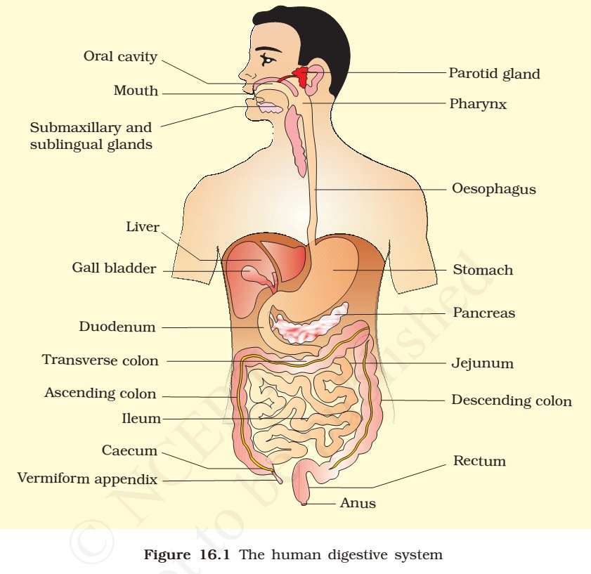
Buccal Cavity or Oral Cavity – Teeth, Tongue, Saliva
- The process of taking food into the body is called ingestion. Ingestion happens through mouth. The mouth leads to the buccal cavity or oral cavity.
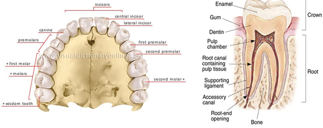
- The oral cavity has a number of teeth and a muscular tongue. Each tooth is embedded in a socket of jaw bone.
- Majority of mammals including human being forms two sets of teeth during their life, a set of temporary milk or deciduous teeth [milk teeth] replaced by a set of permanent or adult teeth [permanent teeth].
- An adult human has 32 permanent teeth which are of four different types, namely, incisors (I), canine (C), premolars (PM) & molars (M).
- Arrangement of teeth in each half of the upper and lower jaw in the order I, C, PM, M is represented by a dental formula which in human is 2123/2123 [2-I,1-C,2-PM,3-M]
- The hard chewing surface of the teeth, made up of enamel (Enamel is the hardest substance in the human body and contains the highest percentage of minerals), helps in the mastication (chewing) of food.
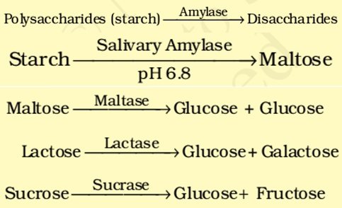
- Our mouth has the salivary glands which secrete saliva. The saliva breaks down the starch into sugars.
- The saliva secreted into the oral cavity contains electrolytes (Na+, K+, Cl”, HCOs) and enzymes, SALIVARY AMYLASE and LYSOZYME.
- The chemical process of digestion is initiated in the oral cavity by the hydrolytic action of the carbohydrate splitting enzyme, the salivary amylase.
- About 30 per cent of starch is hydrolysed here by this enzyme (optimum pH 6.8) into a disaccharide – maltose.
- Lysozyme present in saliva acts as an antibacterial agent that prevents infections.
- The tongue is a fleshy muscular organ attached at the back to the floor of the buccal cavity. It mixes saliva with the food during chewing and helps in swallowing food.
- The tongue is attached to the floor of the oral cavity by the frenulum (a fold of skin beneath the tongue).
- The upper surface of the tongue has small projections called papillae, some of which bear taste buds.
Foodpipe/Oesophagus
- The oral cavity leads into a short pharynx which serves as a common passage for food and air. The esophagus and the trachea (wind pipe) open into the pharynx.
- A cartilaginous flap called epiglottis prevents the entry of food into the glottis during swallowing. [Glottis == opening of the wind pipe].
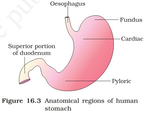
- The swallowed food passes into the foodpipe or oesophagus. The oesophagus is a thin, long tube which extends posteriorly [further back in position] passing through the neck, thorax [the part of the body of a mammal between the neck and the abdomen] and diaphragm [separates the thorax from the abdomen in mammals] and leads to a ‘J’ shaped bag like structure called stomach.
- Mucus in saliva helps in lubricating and adhering the masticated food particles into a bolus. The bolus is then conveyed into the pharynx and then into the oesophagus by swallowing or deglutition.
- The bolus further passes down through the oesophagus by successive waves of muscular contractions called peristalsis. The gastro-oesophageal sphincter controls the passage of food into the stomach.
Stomach
- The inner lining of the stomach secretes mucous, hydrochloric acid and digestive juices.
- The mucous protects the lining of the stomach.
- The acid kills many bacteria that enter along with the food and makes the medium in the stomach acidic.
- The digestive juices break down the proteins into simpler substances.
- A muscular sphincter (gastro-oesophageal) [a ring of muscle surrounding and serving to guard or close an opening] regulates the opening of oesophagus into the stomach.
- The stomach, located in the upper left portion of the abdominal cavity, has three major parts – a cardiac portion into which the oesophagus opens, a fundic region and a pyloric portion which opens into the first part of small intestine.
Small intestine
- Small intestine is distinguishable into three regions, a ‘C’ shaped duodenum, a long coiled middle portion jejunum and a highly coiled ileum.
- The opening of the stomach into the duodenum is guarded by the pyloric sphincter. Ileum opens into the large intestine.
- The small intestine is highly coiled and is about 5 meters long. It receives secretions from the liver and the pancreas. Besides, its wall also secretes juices.
- The digested food passes into the blood vessels in the wall of the intestine. This process is called absorption.
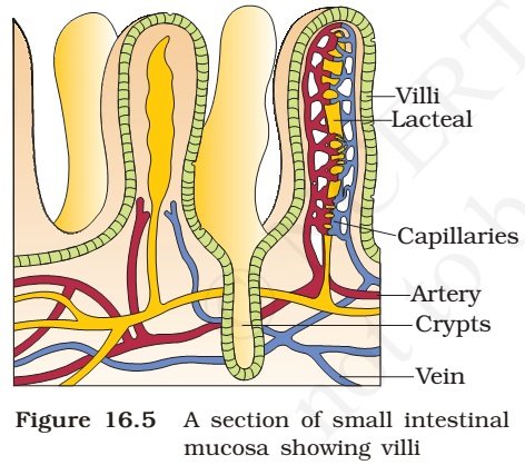
- The inner walls of the small intestine have thousands of finger-like outgrowths. These are called villi (singular villus). The villi increase the surface area for absorption of the digested food.
- Villi are supplied with a network of capillaries and a large lymph (a colourless fluid containing white blood cells) vessel called the lacteal.
- The absorbed substances are transported via the blood vessels to different organs of the body where they are used to build complex substances such as the proteins required by the body. This is called assimilation.
- In the cells, glucose breaks down with the help of oxygen into carbon dioxide and water, and energy is released.
- The food that remains undigested and unabsorbed then enters into the large intestine.
Large intestine
- The large intestine is wider and shorter than small intestine. It is about 1.5 metre in length. Its function is to absorb water and some salts from the undigested food material.
- The remaining waste passes into the rectum and remains there as semi-solid faeces. The faecal matter is removed through the anus from time-to-time. This is called egestion.
Ingestion → Digestion → Absorption → Assimilation → Egestion
- It consists of caecum, colon and rectum. Caecum is a small blind sac which hosts some symbiotic micro-organisms.
- A narrow finger -like tubular projection, the vermiform appendix which is a vestigial organ [small remnant of something that was once more noticeable], arises from the caecum.
| Appendix was helpful in digesting roughage (fibrous indigestible material in vegetable foodstuffs which aids the passage of food and waste products through the gut). Thousands of years ago, when man used to eat roots, leaves, etc., it was essential. But now it has lost its significance. |
- The caecum opens into the colon. The colon is divided into three parts – an ascending, a transverse and a descending part. The descending part opens into the rectum which opens out through the anus.
- No significant digestive activity occurs in the large intestine. The functions of large intestine are: absorption of some water, minerals and certain drugs; secretion of mucus which helps in adhering the waste (undigested) particles together and lubricating it for an easy passage.
- The undigested, unabsorbed substances called faeces enters into the caecum of the large intestine through ileo-caecal valve, which prevents the back flow of the faecal matter. It is temporarily stored in the rectum till defaecation.
Layers of Alimentary Canal
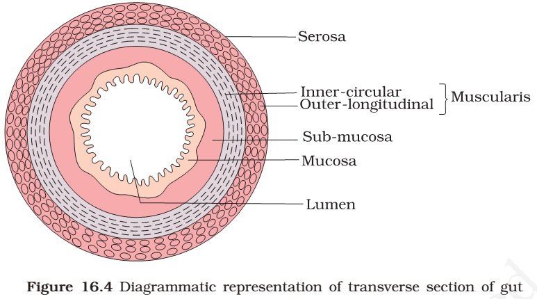
- The wall of alimentary canal from oesophagus to rectum possesses four layers namely serosa, muscularis, sub-mucosa and mucosa.
- Serosa is the outermost layer and is made up of a thin mesothelium (epithelium of visceral organs) with some connective tissues.
- Muscularis is formed by smooth muscles.
- The submucosal layer is formed of loose connective tissues containing nerves, blood and lymph vessels. In duodenum, glands are also present in sub-mucosa.
- The innermost layer lining the lumen of the alimentary canal is the mucosa. This layer forms irregular folds (rugae) in the stomach and small finger-like foldings called villi in the small intestine. Mucosal epithelium has goblet cells which secrete mucus that help in lubrication. Mucosa also forms glands in the stomach (gastric glands).
Digestive Glands
- The digestive glands associated with the alimentary canal include the salivary glands, the liver and the pancreas.
Salivary glands
- Saliva is mainly produced by three pairs of salivary glands, the parotids (cheek), the sub-maxillary (lower jaw) and the sub-linguals (below the tongue).
- These glands situated just outside the buccal cavity secrete salivary juice into the buccal cavity.
- The saliva breaks down the starch into sugars.
Liver
- The liver is a reddish brown gland situated in the upper part of the abdomen on the right side.
- It is the largest gland in the body.
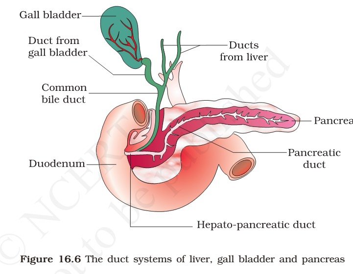
- It secretes bile juice that is stored in a sac called the gall bladder.
- The bile plays an important role in the digestion of fats.
- It has two lobes. The hepatic lobules are the structural and functional units of liver containing hepatic cells.
- The bile secreted by the hepatic cells passes through the hepatic ducts and is stored and concentrated in a thin muscular sac called the gall bladder.
- The duct of gall bladder (cystic duct) along with the hepatic duct from the liver, forms the common bile duct.
- The bile duct and the pancreatic duct open together into the duodenum as the common hepato-pancreatic duct which is guarded by a sphincter called the sphincter of Oddi.
Pancreas
- The pancreas is a large cream coloured gland located just below the stomach.
- The pancreatic juice acts on carbohydrates and proteins and changes them into simpler forms.
- The partly digested food now reaches the lower part of the small intestine where the intestinal juice [succus entericus] completes the digestion of all components of the food.
- The pancreas is a compound (both exocrine and endocrine) elongated organ situated between the limbs of the ‘U’ shaped duodenum.
- The exocrine portion secretes an alkaline pancreatic juice containing enzymes and the endocrine portion secretes hormones, insulin and glucagon.
Digestion – Enzyme Action in Stomach
- The stomach stores the food for 4-5 hours. The food mixes thoroughly with the acidic gastric juice of the stomach by the churning movements of its muscular wall and is called the chyme.
- The proenzyme [inactive precursor of an enzyme] pepsinogen, on exposure to hydrochloric acid gets converted into the active enzyme PEPSIN, the proteolytic (breakdown of proteins or peptides into amino acids) enzyme of the stomach.
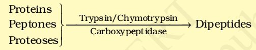
- Pepsin converts proteins into proteoses and peptones (peptides).
- The mucus and bicarbonates present in the gastric juice play an important role in lubrication and protection of the mucosal epithelium from excoriation by the highly concentrated hydrochloric acid. HCl provides the acidic pH (pH 1.8) optimal for pepsins.
- Rennin is a proteolytic enzyme found in gastric juice of infants which helps in the digestion of milk proteins.
Digestion – Enzyme Action in Small Intestine
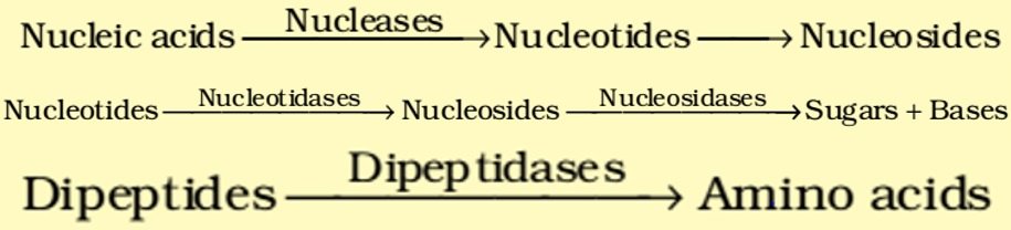
- The pancreatic juice contains inactive enzymes – trypsinogen, chymotrypsinogen, procarboxypeptidases, amylases, lipases and nucleases.
- Trypsinogen is activated by an enzyme, enterokinase, secreted by the intestinal mucosa into active TRYPSIN, which in turn activates the other enzymes in the pancreatic juice.
- The bile released into the duodenum contains bile pigments (bilirubin and biliverdin), bile salts, cholesterol and phospholipids but no enzymes.
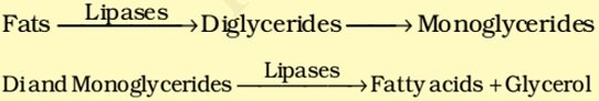
- Bile helps in emulsification of fats, i.e., breaking down of the fats into very small micelles. Bile also activates LIPASES. Small amounts of lipases are secreted by gastric glands.
- The intestinal mucosal epithelium has goblet cells which secrete mucus. The secretions of the mucosa along with the secretions of the goblet cells constitute the intestinal juice.
- This juice contains a variety of enzymes like disaccharidases (e.g., maltase), dipeptidases, lipases, nucleosidases, etc. Hormonal control of the secretion of digestive juices is carried out by local hormones produced by the gastric and intestinal mucosa.
- The mucus along with the bicarbonates from the pancreas protects the intestinal mucosa from acid as well as provide an alkaline medium (pH 7.8) for enzymatic activities.
- The breakdown of biomacromolecules mentioned above occurs in the duodenum region of the small intestine.
- The simple substances thus formed are absorbed in the jejunum and ileum regions of the small intestine.
- The undigested and unabsorbed substances are passed on to the large intestine.
Absorption of Digested Products
- Absorption is the process by which the end products of digestion pass through the intestinal mucosa into the blood or lymph.
- Small amounts of monosaccharides like glucose, amino acids and some electrolytes like chloride ions are generally absorbed by simple diffusion. The passage of these substances into the blood depends upon the concentration gradients.
- However, sometimes substances like glucose and amino acids are absorbed with the help of carrier proteins. This mechanism is called the facilitated transport.
- Transport of water depends upon the osmotic gradient. Active transport occurs against the concentration gradient and hence requires energy. Various nutrients like amino acids, monosaccharides like glucose, electrolytes like Na+ are absorbed into the blood by this mechanism.
- Fatty acids and glycerol being insoluble, cannot be absorbed into the blood. They are first incorporated into small droplets called micelles which move into the intestinal mucosa. They are re-formed into very small protein coated fat globules called the chylomicrons which are transported into the lymph vessels (lacteals) in the villi. These lymph vessels ultimately release the absorbed substances into the blood stream.
- Absorption of substances takes place in different parts of the alimentary canal, like mouth, stomach, small intestine and large intestine. However, maximum absorption occurs in the small intestine.
Summary of Absorption in Different Parts of Digestive System
Mouth |
Stomach |
Small Intestine |
Large Intestine |
| Certain drugs coming in contact with the mucosa of mouth and lower side of the tongue are absorbed into the blood capillaries lining them. | Absorption of water, simple sugars, and alcohol etc. takes place. | Principal organ for absorption of nutrients. The digestion is completed here and the final products of digestion such as glucose, fructose, fatty acids, glycerol and amino acids are absorbed through the mucosa into the blood stream and lymph. | Absorption of water, some minerals and drugs takes place.
|
- The absorbed substances finally reach the tissues which utilise them for their activities. This process is called assimilation.
- The digestive wastes, solidified into coherent faeces in the rectum initiate a neural reflex causing an urge or desire for its removal. The egestion of faeces to the outside through the anal opening (defaecation) is a voluntary process and is carried out by a mass peristaltic movement.
Disorders of Digestive System
- The inflammation of the intestinal tract is the most common ailment due to bacterial or viral infections.
- The infections are also caused by the parasites of the intestine like tapeworm, roundworm, threadworm, hookworm, pin worm, etc.
- Jaundice: The liver is affected, skin and eyes turn yellow due to the deposit of bile pigments.
- Vomiting: It is the ejection of stomach contents through the mouth. This reflex action is controlled by the vomit centre in the medulla. A feeling of nausea precedes vomiting.
- Diarrhoea: The abnormal frequency of bowel movement and increased liquidity of the faecal discharge is known as diarrhoea. It reduces the absorption of food.
- Constipation: In constipation, the faeces are retained within the rectum as the bowel movements occur irregularly.
- Indigestion: In this condition, the food is not properly digested leading to a feeling of fullness. The causes of indigestion are inadequate enzyme secretion, anxiety, food poisoning, over eating, and spicy food.


sir pls post different types of the soil in the world
Is it required for Civils?
very nice sir..thank u
Thanku sir
It’s just copy paste of class 8 Science NCERT Book.