
Magnetic Resonance Imaging (MRI)
- Context (TH): A look at Magnetic Resonance Imaging (MRI), the significance of the technique and its place in modern medical diagnostics.
- Magnetic Resonance Imaging (MRI) is used to obtain images of soft tissues within the body.
- It is a non-invasive diagnostic procedure widely used to image the brain, the cardiovascular system, the spinal cord and joints, various muscles, the liver, arteries, etc.
- MRI scanners use strong magnetic fields & radio waves to generate images of the organs in the body.
|
Uses of MRI
- Observation and treatment of certain cancers, including prostate and rectal cancer.
- Track neurological conditions, including Alzheimer’s, dementia, epilepsy, and stroke.
- Scan changes in blood flow to infer the way the activity of neurons is changing in the brain (in this form, the technique is called functional MRI).
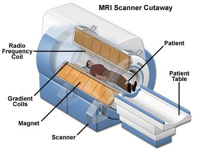
How does MRI work?
- MRI uses a high-power magnet to temporarily change the position of hydrogen atoms, which are abundant in the body’s fat and water.
- As the hydrogen atoms return to their usual position, they emit different amounts of energy depending on the type of tissue they are in. The scanner captures this energy, and a computer creates a picture using this information.
- Unlike x-ray and computed tomography (CT) exams, MRI does not use radiation.
Credits: Deep Medical Centre
Pros
- It can practically image the body from all useful directions and, if required, in very small increments.
- MRI scans don’t pose any threats; once the magnetic fields are taken away, the atoms in the scanned part don’t remain affected.
Cons of MRI
- The magnetic fields that change with time create loud knocking noises, which may harm hearing if adequate ear protection is not used.
- MRI Machines are expensive and are transferred to patients by diagnostic facilities.


